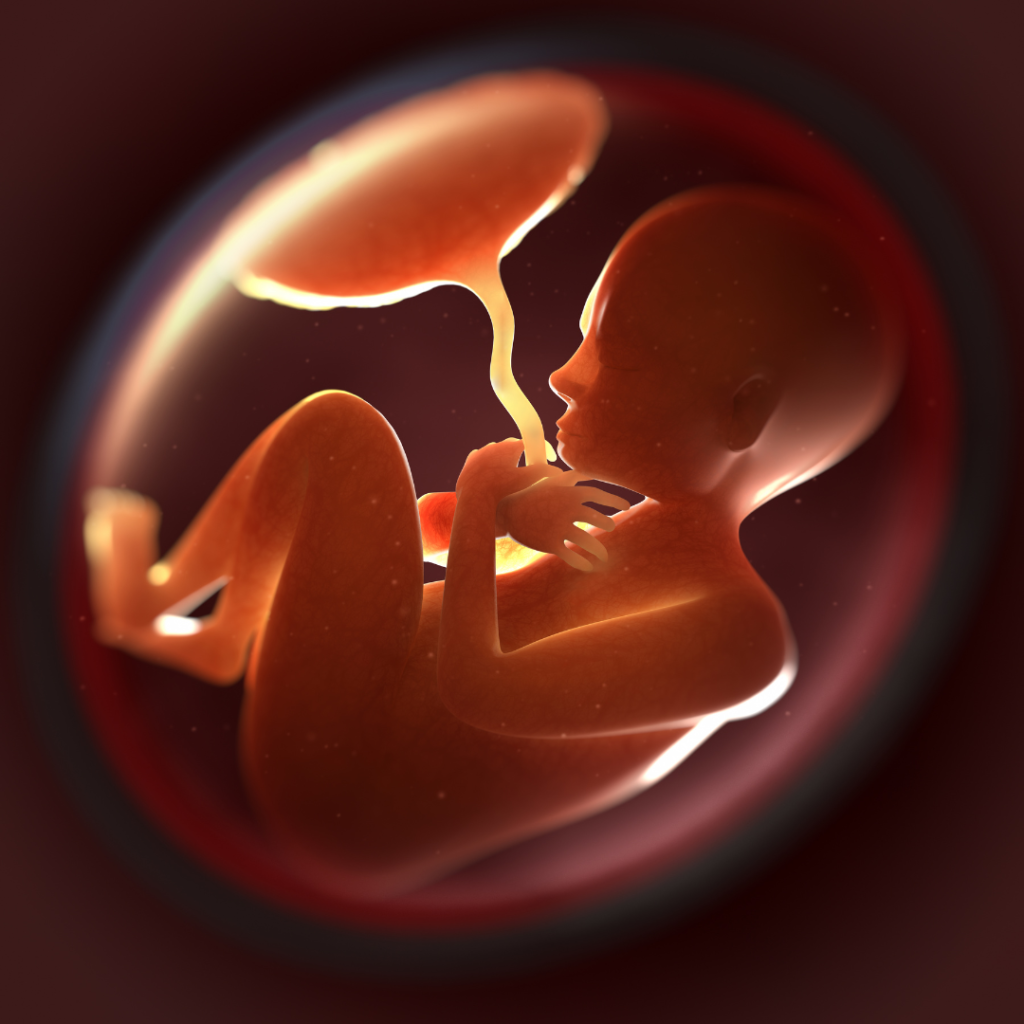
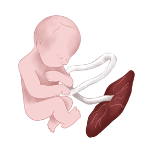
The Placenta is the “Anchor of Life”.
The Placenta is the “Anchor of Life”. Its name derives from the Greek word “plakous/plakounta”, meaning cake due to its round appearance. It is the unique connection between the mother’s womb and her unborn child. Through the placenta, a pregnancy is made possible. It acts as a lung to exchange oxygen and carbon dioxide; as a kidney to filter waste; as a digestive tract to absorb nutrients; as an endocrine gland that produces many hormones and growth factors to ensure a stable hormonal milieu and finally, as an immunological barrier to fend off rejection of the pregnancy. The unique immunological and hormonal environment created in the so called “fetoplacental unit” ensures the undisturbed and timely growth of the child by undisrupted oxygen and nutrient supply to and disposal of waste from the child’s metabolic processes. The two sides (maternal and fetal) never mix but are tangent to each other to ensure continuous exchange. A failure in the placenta endangers the pregnancy.
​
After delivery, the placenta will detach itself and will also be delivered in the so-called afterbirth. The Placenta and its associated Amniotic membranes/tissues are extremely rich in Mesenchymal Stromal Cells (MSC) with huge regenerative potential. MSC from these tissues have a broader differentiation potential than the Cord Tissue (Wharton’s Jelly) MSC and can differentiate to bone, cartilage, heart, fat, muscle, nerve, liver, pancreas, blood vessel, immune system populations and skin among some of the directions they can take. MSC are well tolerated by the host and can therefore avoid allo-recognition by the recipient’s immune system. These cells are able to secrete cytokines and generate a local immunosuppressive microenvironment to reduce inflammation associated with many degenerative conditions. “They offer a renewable source of cell replacement for antiaging therapy, regeneration medicine, and are used to treat various neurological and immune disorders. Placental stromal cell therapy will be a promising answer for many of today’s untreatable diseases in the years to come” (1).
​
​
Table 10.1 Differentiation potential of human placenta
and amniotic fluid mesenchymal stem cells
​
Cell Type
​
Expression marker and/or phenotype
​
Multi-lineage SP cells
​
HLA I(-)/II(-), HLA I(+)/TI(-)
Angiogenic
​
Flt-1, KDR, ICAM-1, CD34
Hepatic
​
Albumin, HNF-4, Al AT
Pancreatic
PDX-1, PAx-6, NKx2.2, insulin, glucagons
Neurogenic
​
Release of catecholamine,
acetylcholine, neurotropic factors,
activin, noggin, neurofilament
Skeletal myogenic
​
Myosin
Cardiomyogenic
​
MyoD, desmin
Adipogenic
​
Oil Red-O stain
Chondrodenic
​
collagen type II immunochemical detection
Osteogenic
​
von Kossa stain, alkaline phosphatase, osteocalcin
*Side populations cells
San Rafael (CA): Morgan & Claypool Life Sciences.
Copyright © 2010 by Morgan & Claypool Life Sciences.
At THAI StemLife,
we can also sometimes offer
collection of Placental Blood.
This can be an option when the cord blood volume collected is low.
​
Placental collection happens “ex-utero”, meaning it will happen when the placenta has been delivered. Placental blood is the same as cord blood and will be processed similarly.
MSC from those sources are different than blood stem cells from the cord blood. Those MSC are more primitive than cord tissue MSC and have broader differentiation capacity as seen in the above table with endothelial stem cells that can promote blood vessel formation, and epithelial stem cells that are precursors to the growth of skin. Amniotic membranes have been for decades used in burns and other skin applications, in orthopedic surgery, dentistry, urology, ENT surgery and neurosurgery while placental tissue has been applied in eye surgery since a long time. Placental vessels are being used as vascular grafts. A growing number of clinical research is ongoing studying the above mentioned MSC in autoimmune diseases, skin applications, orthopedics, etc. At THAI StemLife, you will be offered two options. One will be to freeze and store tissue for future use. The second option comprises the first but also includes culture of MSC from the tissue before freezing both.